Polychromatic Scanning Electron Microscopy
(Color-coded visualization of small- and large-scale variability in surface topography)
Polychromatic Scanning Electron Microscopy
(Color-coded visualization of small- and large-scale variability in surface topography)


3D Printing
Publications
News
Gallery
Contact
Team

Research
Imaging
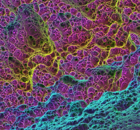

Aluminum ductile fracture
Field diameter: 375 µm
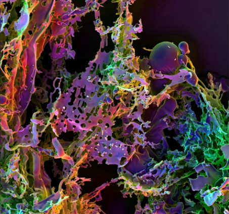
Self-assembling peptides
Field diameter: 750 µm
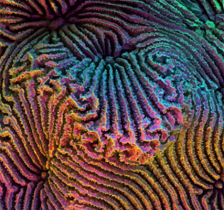
Meandroid coral skeleton
Field diameter: 4 mm
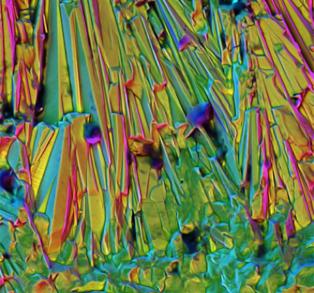
Polycrystalline manganese oxide
Field diameter: 125 µm