Stereo Scanning Electron Microscopy
(Reveling high-resolution three-dimensional material structural complexity)
Stereo Scanning Electron Microscopy
(Reveling high-resolution three-dimensional material structural complexity)


Imaging
3D Printing
Publications
News
Gallery
Contact
Team
Research
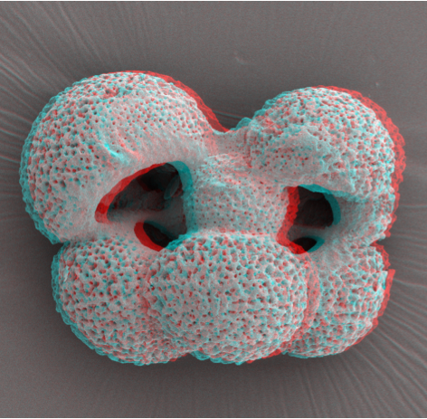
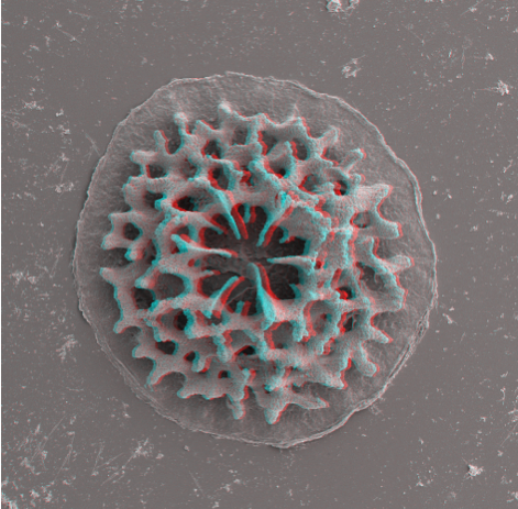
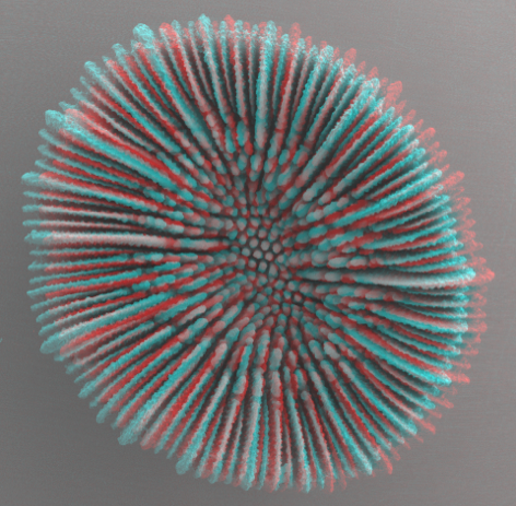
“Twinned” test of a planktonic foraminfera (Globigenina sp.)
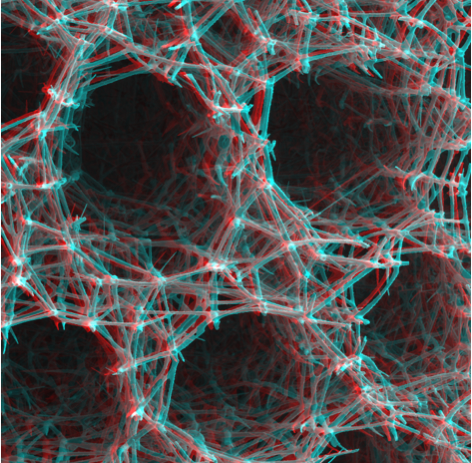
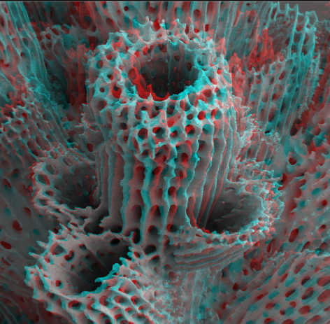
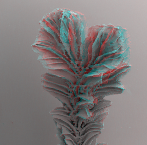
Pacific hexactinellid sponge skeleton (Aphrocallistes sp.)
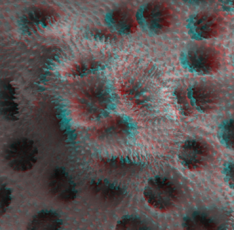
Post-metamorphic Atlantic coral skeleton (Agaricia sp.)
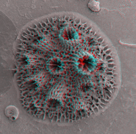
Post-metamorphic Pacific coral skeleton (Acropora sp.)
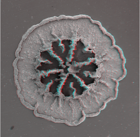
Juvenile Pacific coral skeleton (Acropora sp.)
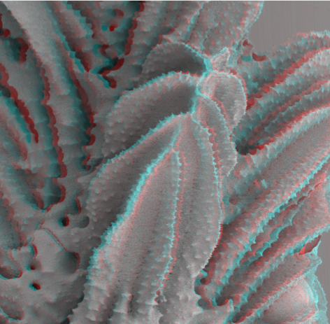
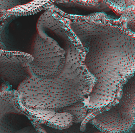
Pacific coral skeleton (Acropora sp.)
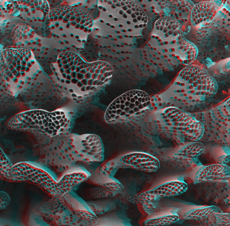
Pacific coral skeleton (Hydnophora sp.)
Pacific coral skeleton (Hydnophora sp.)
Pacific coral skeleton (Pocillopora sp.)
Pacific coral skeleton (Paracyathus sp.)
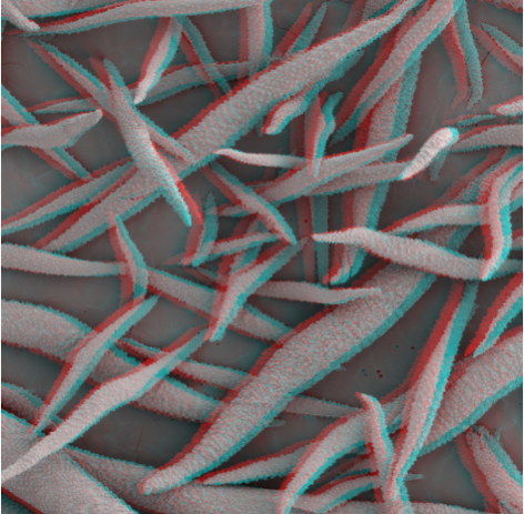
Soft coral sclerites (Sinularia sp.)
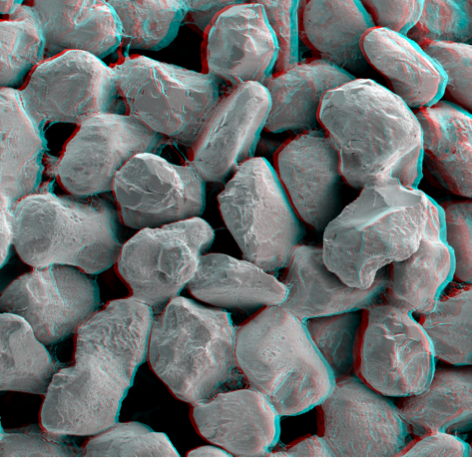
Consolidated sand grains from a Phragmatopoma sp. tube
Bryozoan skeleton (Watersipora sp.)
Bryozoan skeleton (Diaperoecia sp.)
One major drawback to a greatly enhanced depth of field in traditional scanning electron microscopy is the perceived loss of the apparent three dimensionality of an object. As a result, the images can often appear flat and critical topographical features can be obscured. Stereo scanning electron microscopy can help resolve these issues by revealing previously undetectable three dimensional architectural complexities. To create this effect, two SEM images are acquired from different angles and the images are digitally combined to create an incredibly realistic composite.
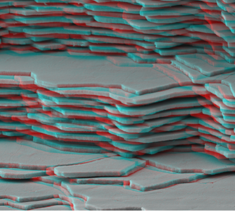
Abalone nacre (Haliotis sp.)